Diagrams of the Female Reproductive System 101 Diagrams
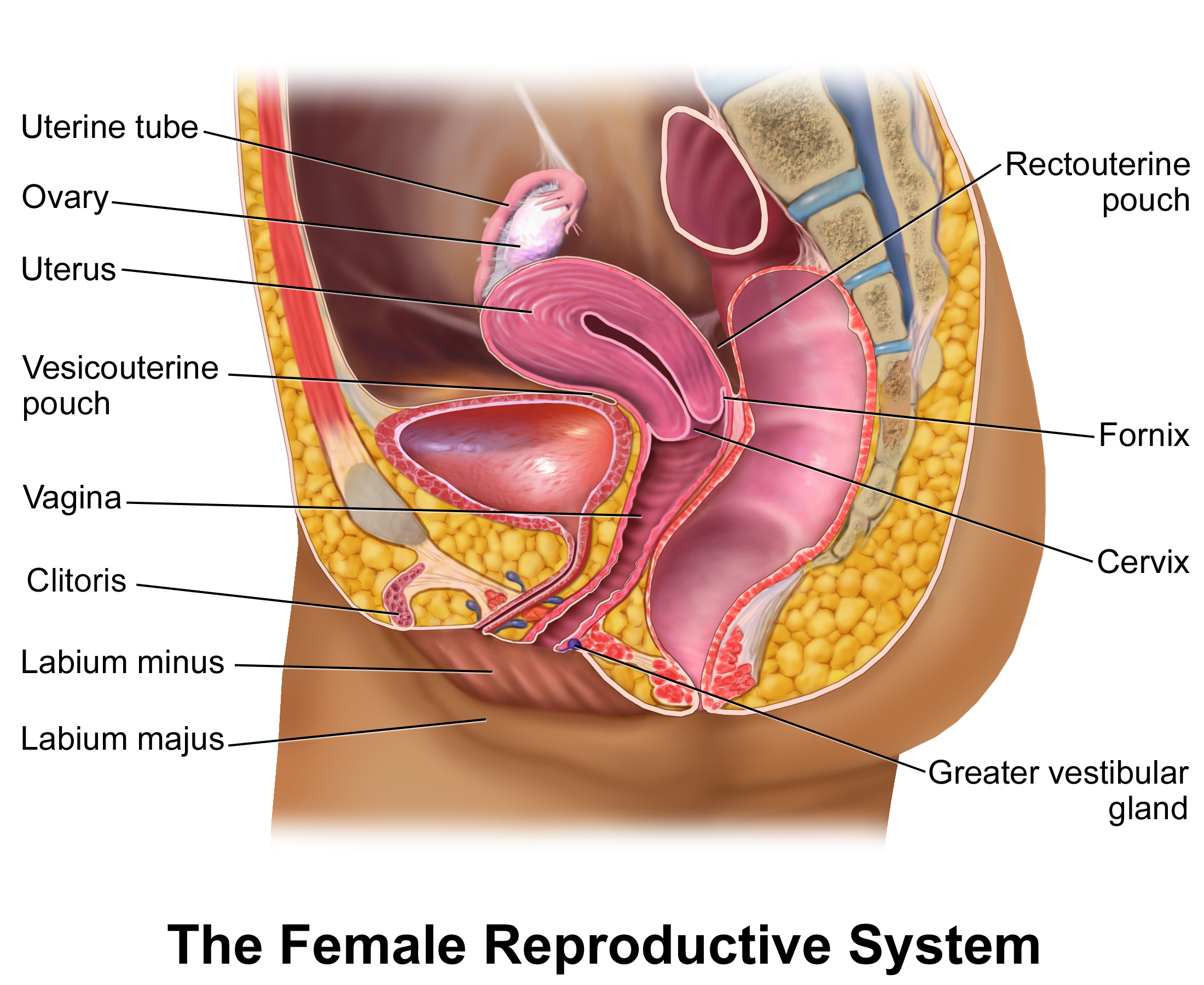
Vagina. The vagina, shown at the bottom of Figure 27.9 and Figure 27.10, is a muscular canal (approximately 10 cm long) that serves as the entrance to the reproductive tract.It also serves as the exit from the uterus during menses and childbirth. The outer walls of the anterior and posterior vagina are formed into longitudinal columns, or ridges, and the superior portion of the vagina—called.
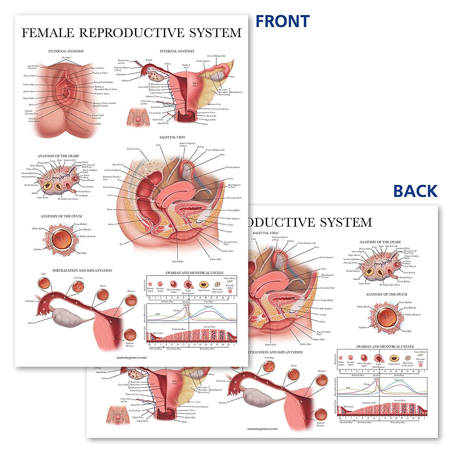
Female Reproductive System Anatomy Posters
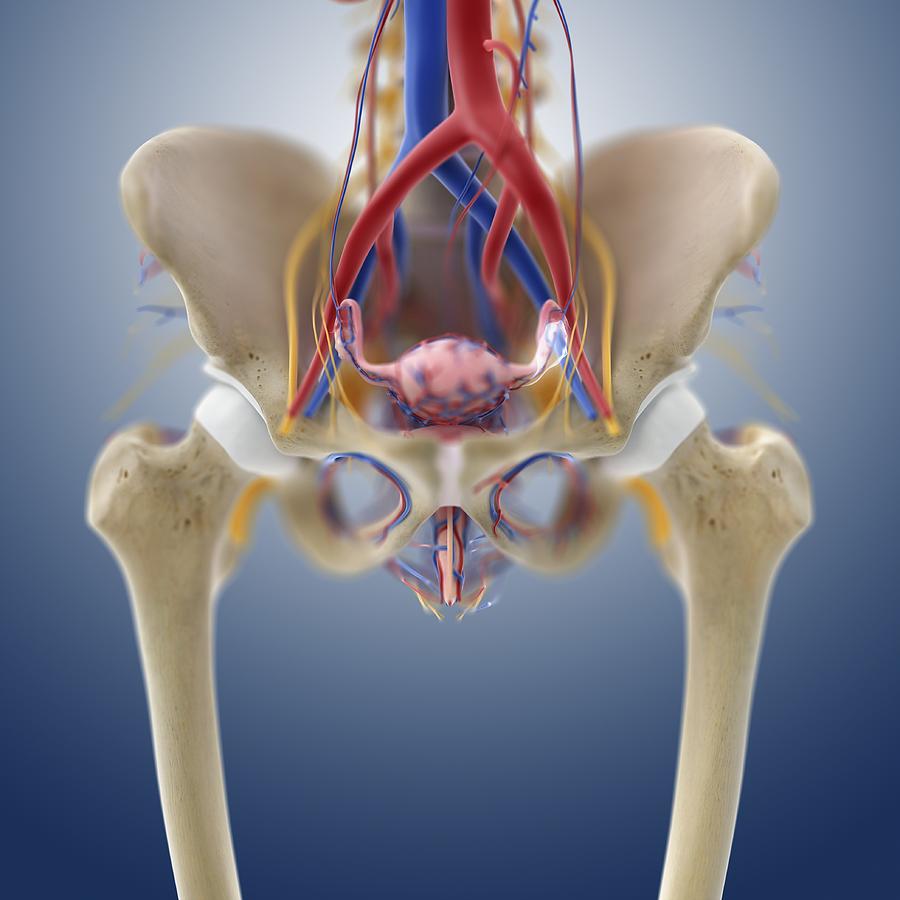
There are a wide variety of indications for imaging the female pelvis. From acute pelvic pain and abnormal uterine bleeding (AUB) to identifying and characterizing pelvic masses or potential causes of infertility, imaging plays a crucial role in diagnosis and management.

Human Reproduction Part 1 Female Reproductive System Leaving Cert Riset
App to train reading breast tomosynthesis images COMMUNITY. e-Cases Imaging clinical cases zoo-Paedia. This e-Anatomy module contains a hundred and one illustrations dedicated to the anatomy of the female pelvis. These fully annotated anatomical illustrations are presented as a comprehensive atlas of the female reproductive system, bladder.
.jpg?response-content-disposition=attachment)
Female Reproductive System resource Imageshare
Browse 4,505 female reproductive system anatomy photos and images available, or search for female anatomy to find more great photos and pictures. Browse Getty Images' premium collection of high-quality, authentic Female Reproductive System Anatomy stock photos, royalty-free images, and pictures.
Female reproductive anatomy, artwork Photograph by Science Photo
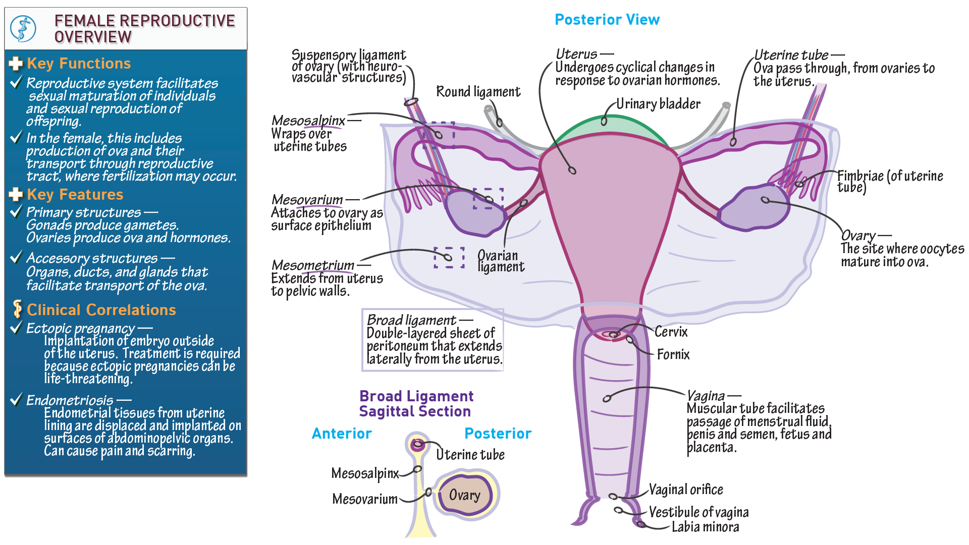
1/5 Synonyms: none The female sex organs consist of both internal and external genitalia. Together they comprise the female reproductive system, supporting sexual and reproductive activities. The external genital organs, or vulva, are held by the female perineum.
The Female Reproductive System Complete Anatomy Gambaran
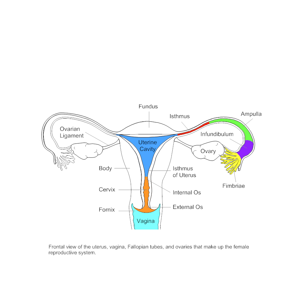
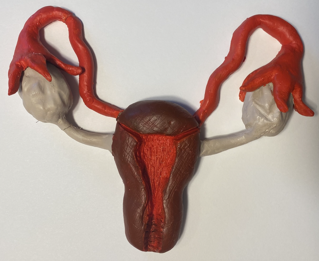
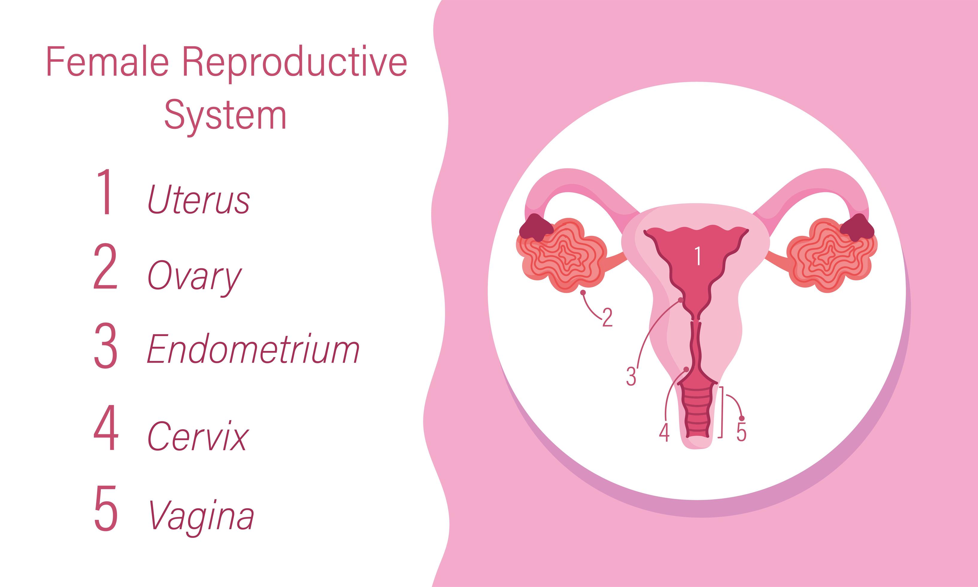
Anatomy of the female reproductive system. The organs in the female reproductive system include the uterus, ovaries, fallopian tubes, cervix, and vagina. The uterus has a muscular outer layer called the myometrium and an inner lining called the endometrium. Normal female reproductive system anatomy. Yes - This image is copyright protected.
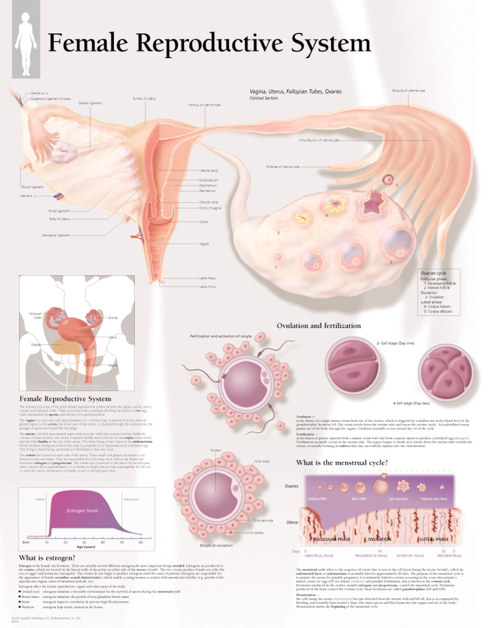
Female Reproductive System Scientific Publishing
Female reproductive system 1. Vulva: 2. Labia majora; 3. Labia minora; 4. Vestibule; 5. Clitoris: (with 6. Glans and 7. Body). 8. Bulb of vestibule 9. Vagina: 10. Hymen; 11. Lumen; 12. Wall; 13. Fornix (lateral) 14. Uterus: Parts: 15. Cervix; 16. Body and 17. Fundus. 18. Orifices: External and Internal; 19. Cervical canal; 20.
The Diagram Of Female Reproductive Organs Diagram Of The Female
Anatomy, Abdomen and Pelvis: Female External Genitalia. Nguyen JD, Duong H. StatPearls. 2023 Jan Normal vulvovaginal, perineal, and pelvic anatomy with reconstructive considerations.
Reproductive System Female External
Episode 1: Meet Your Vagina & Vulva | Planned Parenthood Video Sexual anatomy that's typically called female includes the vulva and internal reproductive organs like the uterus and ovaries What are the external parts? The vulva is the part of your genitals on the outside of your body.

Section of female reproductive system Female reproductive system
Interactive Guide to Female Reproductive Anatomy | Innerbody The Female Reproductive System 3D models, illustrations, and accompanying descriptions guide you in exploring the anatomy and physiology of the female reproductive organs. Learn all about fertilization, pregnancy, delivery, and lactation. By: Tim Taylor Last Updated: Feb 15, 2022
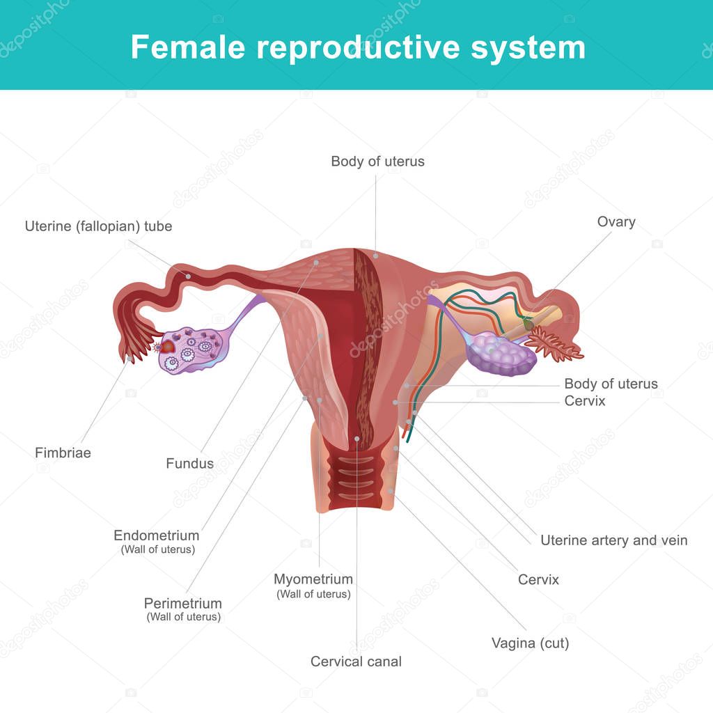
Diagram Of Internal Organs Female Female Reproductive System Internal
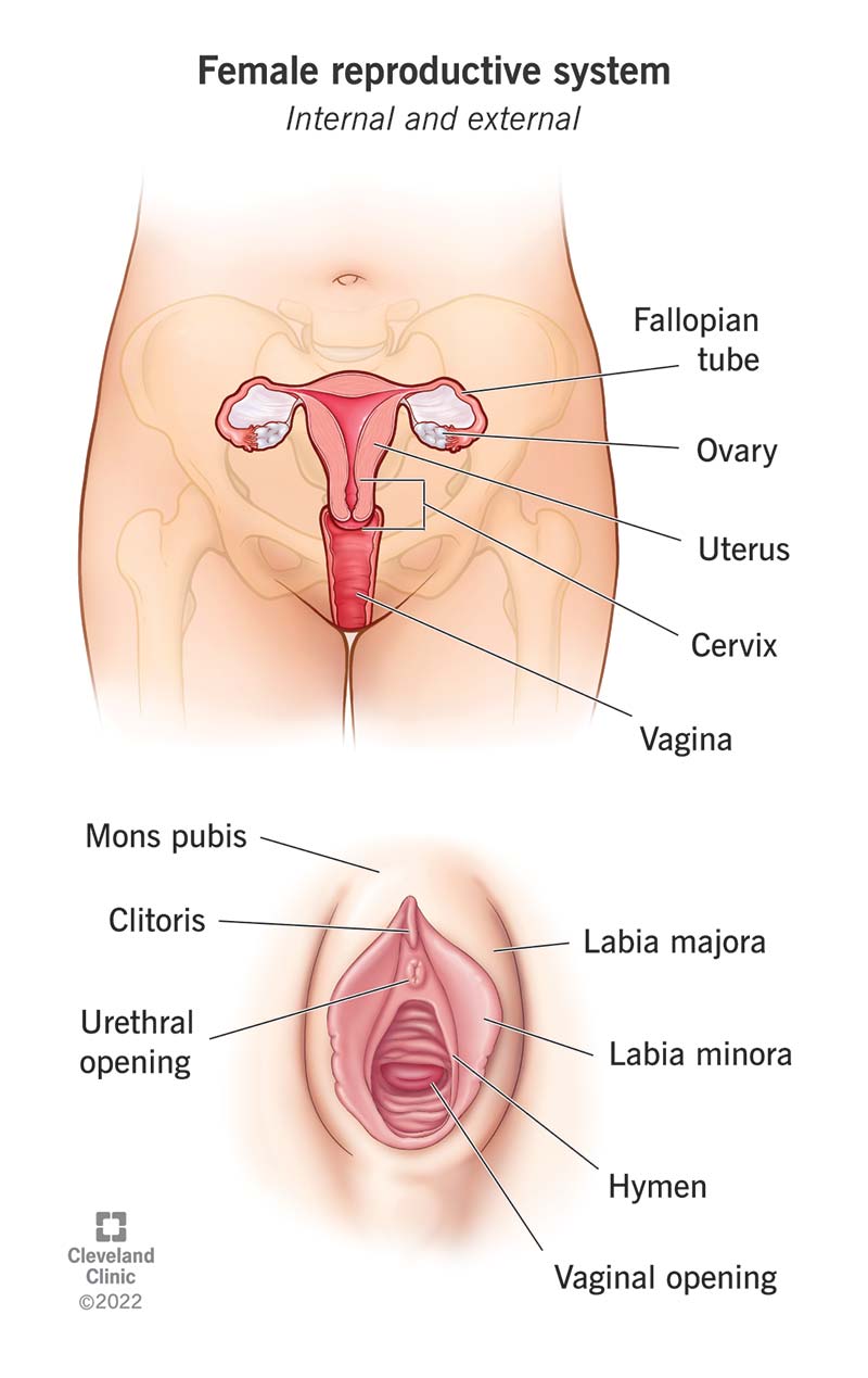
The external female genitalia are a part of the female reproductive system, and include the: mons pubis, labia majora, labia minora, clitoris, vestibule, hymen, vestibular bulb and vestibular glands. The components of the external female genitalia occupy a large part of the female perineum and collectively form what's known as the vulva.

The female reproductive system Complete Anatomy
Browse 15,129 authentic female reproductive organ stock photos, high-res images, and pictures, or explore additional gynecological examination or uterus stock images to find the right photo at the right size and resolution for your project. Related searches: gynecological examination uterus female anatomy anatomy ovary NEXT

Female Reproductive Anatomy Reproductive Medbullets Step 1
The vagina is part of the internal genitalia of the female reproductive system. The internal female sex organs form a pathway, the internal female genital tract, composed of the vagina, uterus, the paired uterine tubes and ovaries. The vagina serves a multitude of functions. It facilitates menstruation, childbirth and sexual intercourse, as it.

Detailed Female Reproductive System Medical Edition 3D model CGTrader
The female reproductive anatomy includes both external and internal parts. External parts. The function of your external genitals are to protect the internal parts from infection and allow sperm to enter your vagina. Your vulva is the collective name for all your external genitals. A lot of people mistakenly use the term "vagina" to.
Anatomy Picture Of Female Reproductive System Detailed Female
Browse 3,096 authentic reproductive anatomy stock photos, high-res images, and pictures, or explore additional female reproductive anatomy or male reproductive anatomy stock images to find the right photo at the right size and resolution for your project. female reproductive anatomy male reproductive anatomy male reproductive anatomy illustration
female human reproductive system diagram of the internal organ 2777494
The female reproductive organs can be subdivided into the internal and external genitalia (see the images below). The internal genitalia are those organs that are within the true pelvis. These include the vagina, uterus, cervix, uterine tubes (oviducts or fallopian tubes), and ovaries. The external genitalia lie outside the true pelvis.